Fig. 3.
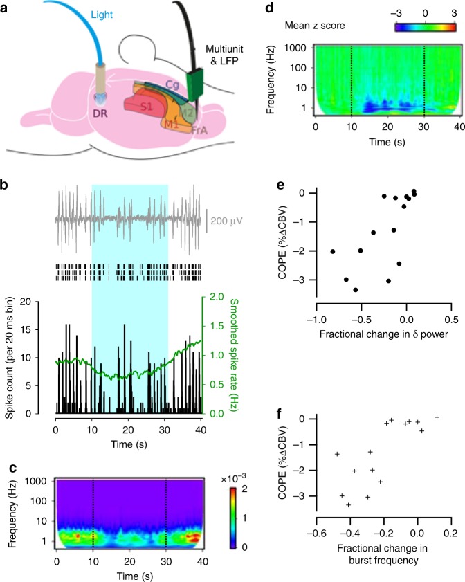
Photoactivation of DRN 5-HT neurons dampens MUA and LFP power in cortical projection areas. a Schematic of the experimental set-up for paired MUA and LFP recordings in the DRN and projection areas during photostimulation. b Cortical LFP (top), raster plots (middle) of spikes recorded on 3 neighbouring channels and spike time histogram (bottom), reveal reduction in the frequency of cortical network bursts and MUA during optogenetic activation of DRN 5-HT neurons. c Wavelet amplitude spectrum of the cortical LFP averaged over 6 stimulation trials for the experiment shown in (b) (warmer colours indicate higher power). Dashed lines indicate onset and end of photostimulation. d Z transform of the wavelet amplitude spectrum averaged across recording sites of mice with robust CBV response to photostimulation, showing a consistent reduction in the amplitude of low-frequency LFP components (8 recording sites from 3 mice). e, f COPEs from various cortical regions correlate strongly with fractional changes in both the d delta power (Pearson r = 0.69; p = 0.004) and e burst frequency (Pearson r = 0.78; p = 0.0006) recorded in 15 recording sites from 7 mice

