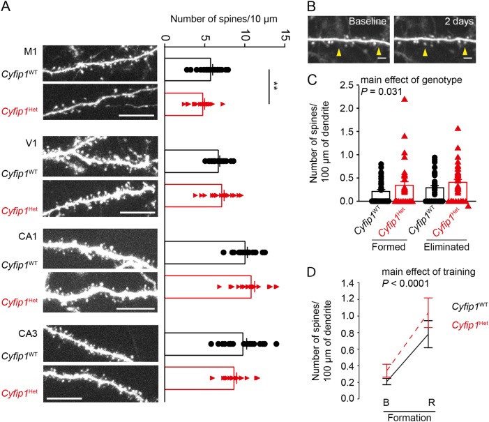
Fig. 3. Cyfip1 haploinsufficiency leads to decreased stability of dendritic spines without affecting their formation.
a Dendrites of GFP-expressing neurons from the motor cortex (M1), visual cortex (V1), and hippocampus (CA1 and CA3) of Cyfip1WT and Cyfip1HET mice. Scale bars represent 10 μm. Quantification of spine density of Cyfip1WT and Cyfip1HET dendrites. b Two images of the same dendrite of a Cyfip1WT mice motor neuron taken at an interval of 2 days showing the newly formed dendritic spines (arrows). Scale bars represent 2 μm. c Quantification of formed and eliminated dendritic spines in motor cortices of Cyfip1WT and Cyfip1HET mice. d Quantification of formed dendritic spines in motor cortices of Cyfip1WT and Cyfip1HET mice before (B) and after rotarod training (R). All values are represented as mean ± SEM. In (b) and (d), statistical significance was tested by ANOVA followed by Bonferroni post-hoc test. **P ≤ 0.01

