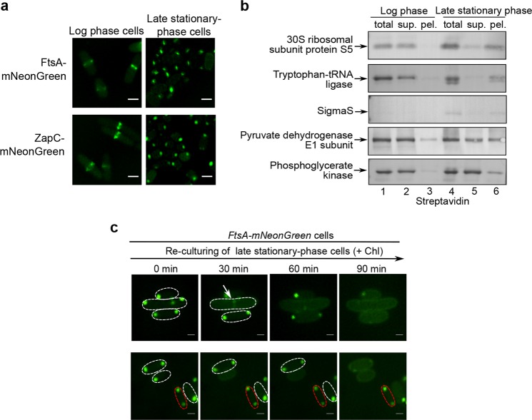
Fig. 4. Regrowth-delay bodies selectively sequester multiple key proteins that are released to re-function when cells exit their regrowth lag and resume growth.
a Fluorescence microscopic images of the log-phase (left) and late stationary-phase (right) E. coli cells in which mNeonGreen-fused FtsA or ZapC (both being identified in the regrowth-delay bodies by mass spectrometry analysis, as shown in Supplementary Fig. S9a) was expressed from a plasmid under the control of a constitutive promoter. Scale bars, 1 μm. b Blotting results to analyze the indicated Avi-tagged proteins in the indicated lysate fractions of the log-phase or late stationary-phase wild-type cells, probed with streptavidin-AP. c Fluorescence microscopic images of the re-cultured late stationary-phase cells in which FtsA-mNeonGreen was expressed from a plasmid under the control of a constitutive promoter in fresh medium containing chloramphenicol, obtained at the indicated time points. Scale bars, 1 μm

