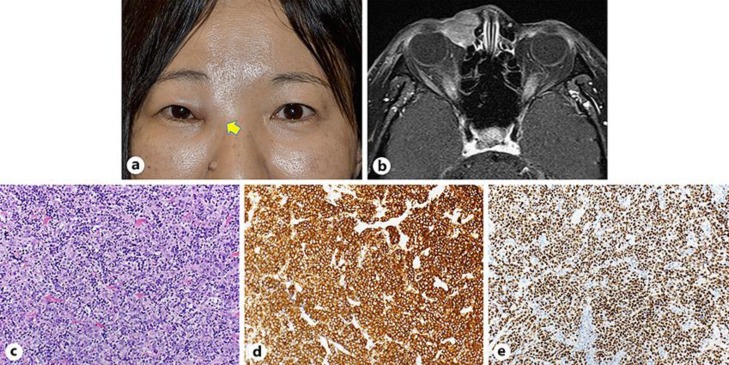
Fig. 1.
a The patient's photograph during the first examination showing a mass in the right medial canthus. b Enhanced T1-weighted axial magnetic resonance imaging showed a soft tissue mass in the lacrimal sac with heterogeneously mild enhancement. c–e Histopathological and immunostaining specimens from the lacrimal sac. c Diffuse proliferation of large lymphoid cells with high-density nuclei (hematoxylin and eosin, ×200). Immunostaining with CD20 (d) and BCL-6 (e) were both positive (×200).

