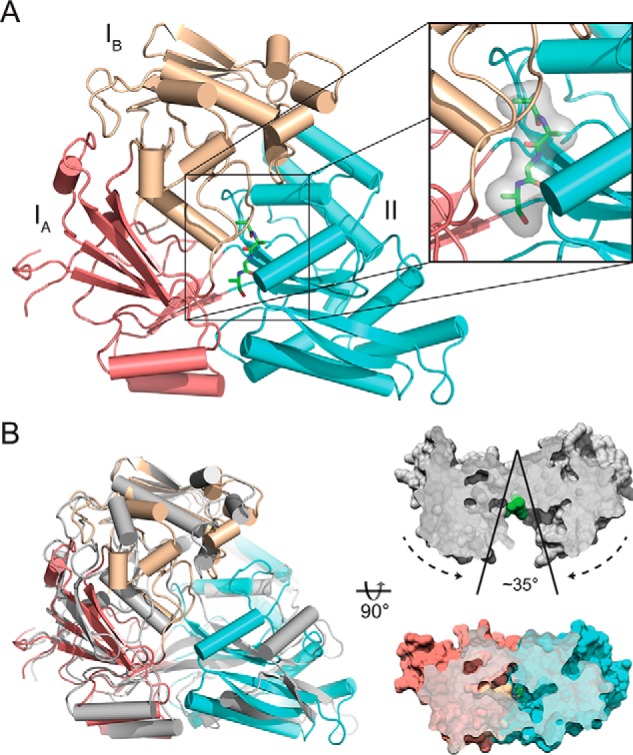
Figure 1.

Crystal structure of nthiOppA in closed ligand-bound conformation. A, crystal structure of nthiOppA, colored by domain (IA, salmon; IB, wheat; II, cyan), bound to co-purified peptide in green. Inset, surface and stick representation of the co-purified peptide in substrate-binding pocket. B, cartoon representation of the ligand-bound structure of nthiOppA aligned with the open unbound ecOppA structure in gray (PDB code 3TCH), and surface representation of cross-sections of ecOppA and nthiOppA. Based on the alignment with nthiOppA, the green spheres in the open unbound ecOppA structure highlight the binding site where peptide interacts with the SBP to form the closed conformation.
