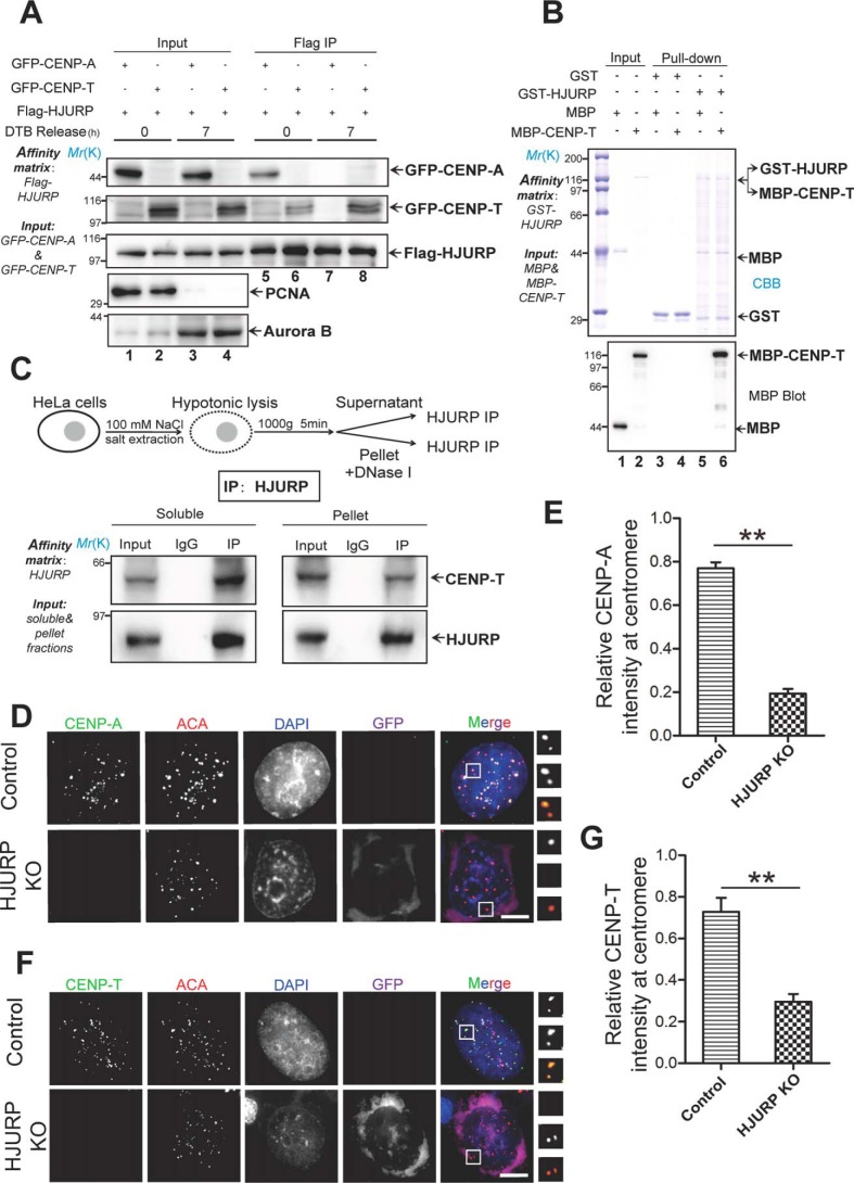
Figure 1.
HJURP interacts with and specifies CENP-T localization to the centromere. A, aliquots of HeLa cells were transiently transfected to express FLAG-HJURP with GFP–CENP-T or GFP–CENP-A. 4 h after transfection, the cells were synchronized by a double thymidine block (DTB) protocol, and synchronized cells were released into G1/S phase (0 h) or G2 phase (7 h). Then the synchronized cells were lysed, and clarified cell lysates were incubated with FLAG-agarose. The beads were washed and boiled in 1× SDS-PAGE sample buffer prior to electrophoresis. Results were analyzed by Western blotting with the indicated antibodies. Each blot was cut around the expected band and is presented with molecular weight information. PCNA, proliferating cell nuclear antigen. B, HJURP interacts directly with CENP-T in vitro. GST-tagged HJURP recombinant protein bound on GSH-agarose beads was used as an affinity matrix to isolate MBP–CENP-T purified from bacteria. Results were analyzed by anti-MBP blot. C, top panel, schematic of the experimental preparation of cell extract. Synchronized HeLa cells were lysed at various hypotonic NaCl concentrations, and the supernatant was separated by centrifugation (1,000 × g, 5 min). The remaining fraction was further treated with DNase I. Bottom panel, HJURP antibody and IgG were incubated with supernatant and pellet fractions for 1 h, and then protein A/G was added to the incubation for another 1 h. Western blotting showed that HJURP interacts with CENP-T in both the supernatant and pellet in vivo. IP, immunoprecipitation. D, HJURP KO plasmid pools were transfected into HeLa cells. Seventy-two hours after transfection, HeLa cells were fixed, followed by a standardized immunofluorescence staining protocol for CENP-A (green), ACA (red), GFP (purple), and DNA (blue). Representative images were collected from three independent experiments and are presented. Scale bar = 5 μm. E, statistical analyses of CENP-A centromere localization level under efficient HJURP knockout. Over 25 cells were tested in each category from three independent preparations. Data represent mean ± S.E. Statistical significance was tested by two-sided t test. **, p < 0.01. F, HJURP KO plasmids were transfected into HeLa cells, and centromere localization of CENP-T was analyzed using the indicated antibodies. As indicated, HJURP has a critical role in CENP-T centromere localization. CENP-T, green; ACA, red; GFP, purple; DNA, blue. Scale bar = 5 μm. G, statistical analysis of CENP-T centromere localization level upon HJURP knockout. Over 25 cells were tested in each category from three independent preparations. Data represent mean ± S.E. Statistical significance was tested by two-sided t test. **, p < 0.01.

