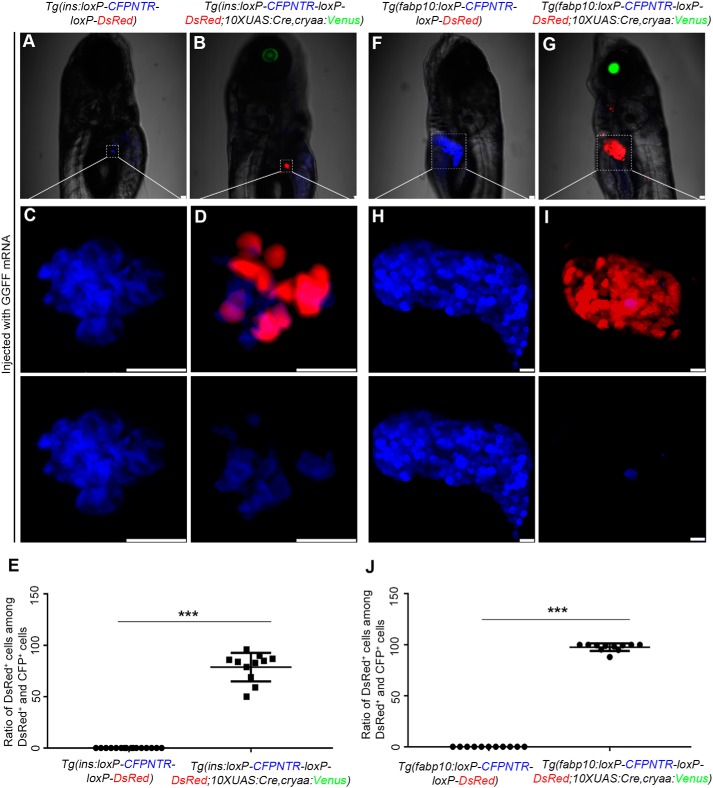
Figure 2.
The validation of the Cre/loxP–based transgenic reporter lines. A–D, the fluorescence of the pancreatic β cells in Tg(ins:loxP-CFPNTR-loxP-DsRed; 10×UAS:Cre, cryaa:Venus) was converted from blue to red by GGFF mRNA. E, quantification of the percentage of the DsRed+ cells among the DsRed+ and CFP+ cells in C and D. F–I, the color of the hepatocytes of the Tg(fabp10:loxP-CFPNTR-loxP-DsRed; 10×UAS:Cre, cryaa:Venus) shifts from blue to red when injected with GGFF mRNA at one-cell stage. J, quantification of the percentage of the DsRed+ cells among the DsRed+ and CFP+ cells in H and I. Asterisks indicate statistical significance: ***, p < 0.001. Scale bars, 20 μm. Error bars, ±S.D.

