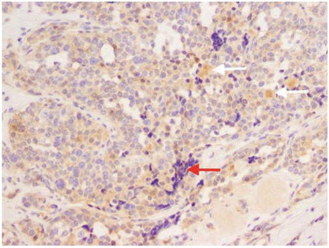
Figure 3.
Faint staining of the RASAL1 protein in the IHC analysis in the medullar thyroid carcinoma tissue (20 ×). Normally the RASAL1 gene is very highly expressed in thyroid cells since the thyroid along with the brain and adrenal glands are the sites of highest expression. White arrow: Cluster of cells that are positive but that do not look like tumor cells. Red arrow: Lymph nodes: a negative control.

