Figure 1.

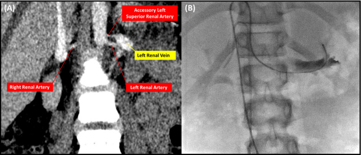
Findings from computed tomographic angiography of the abdomen showing small accessory left superior renal artery (A) and renal venous catheter with its tip placed in the draining vein of the superior pole of the left kidney (B).

Findings from computed tomographic angiography of the abdomen showing small accessory left superior renal artery (A) and renal venous catheter with its tip placed in the draining vein of the superior pole of the left kidney (B).