Fig. 2.
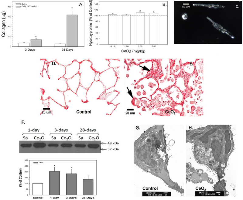
Cerium oxide exposure induced pulmonary fibrosis. Soluble collagen in the first BALF (A) and hydroxyproline content in lung tissue (B) from control and cerium oxide (3.5 mg/kg)-exposed rats. Illuminated CeO2 particles, using dark field-based illumination, were clearly detected in fibroblasts from particle-exposed lungs (C), but not in controls. Light micrograph of sirius red staining for collagen formation in the control (D) and CeO2-exposed (E) lung tissue at 28 days post-exposure. Arrows indicate increased collagen (scale bar = 20 μm). Immunoblot analysis of α-SMA in control or CeO2-exposed lung tissues (F) at different time points after exposure. TEM micrographs of control (G) and CeO2-exposed (H) lung tissue at 28 days post-exposure (bar = 1 μm). *Significantly different from control at p < 0.05.

