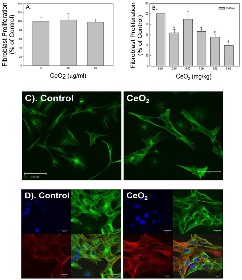
Fig. 3.
Effects of cerium oxide exposure fibroblast function. The proliferation rate of naïve primary fibroblasts in response to CeO2 exposure in vitro (A) and fibroblasts collected from control and CeO2-exposed rats (B). Immunofluorescence staining of fibroblasts for α-tubulin (green) obtained from control and CeO2-exposed rats at 28 days post exposure (bar = 100 μm) (C). Immunofluorescence for α-SMA (red) in α-tubulin stained fibroblast (green) isolated from control and CeO2-exposed rats at 28 days post exposure. DAPI (blue) was used as nuclei marker (bar = 20 μm) (D).

