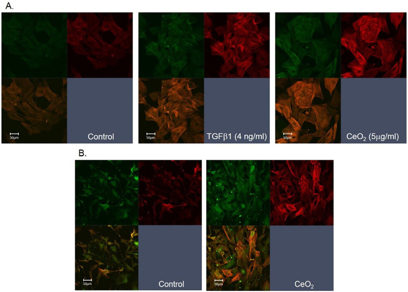
Fig. 5.
In vitro and in vivo CeO2 exposure-induced EMT in ATII cells. (A) In vitro exposure of naïve ATII cells to CeO2 or TGF-β1 induced EMT. Myofibroblast formation was monitored using α-SMA staining of the formation of spindle like fibers (red). The epithelial cells were stained with pan-cytokeratin (green). (B) Confocal micrographs of ATII cells harvested from control or CeO2 (3.5 mg/kg)-exposed rats at 3 days post exposure.

