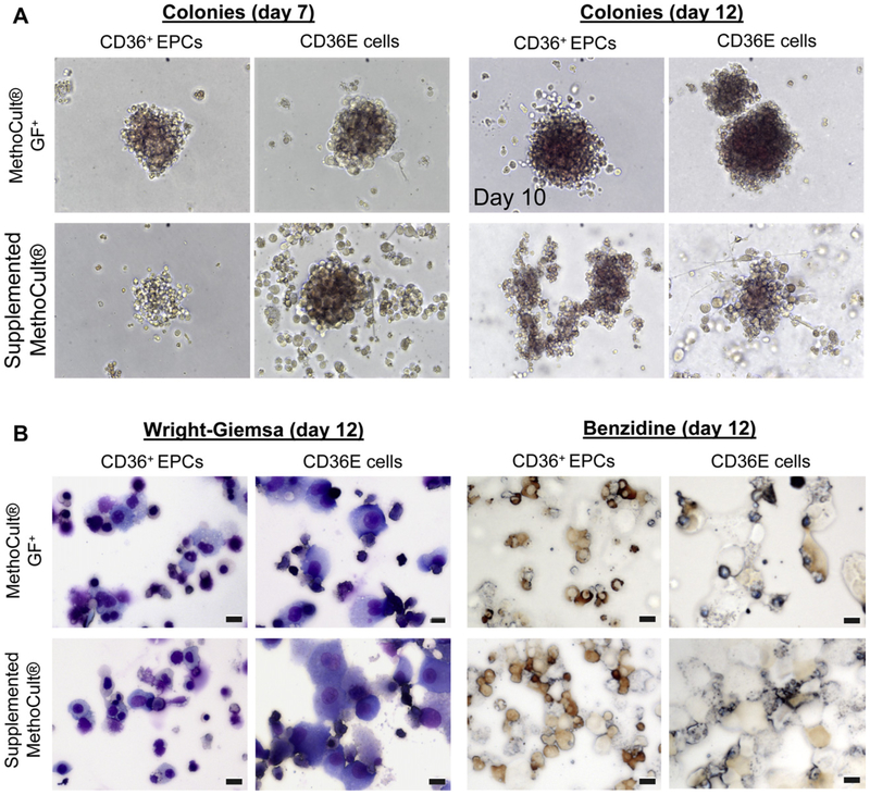
Figure 4.
Colony-forming cell assay of CD36E cells. CD36E cells were cultured in two different methylcellulose-based media: the MethoCult GF+ for various kinds of hematopoietic colony formation; the supplemented MethoCult for erythroid colony formation. (A) Using a light microscope at 200× magnification, colonies were evaluated on days 7 and 12, but colonies from CD36+ EPCs cultured in the MethoCult GF+ were evaluated on day 10, instead of day 12, due to overgrowth. (B) On day 12, individual colony cultures were harvested, cytocentrifuged onto glass slides, and stained with Wright-Giemsa or benzidine followed by microscopy. Scale bars: 10 μm.

