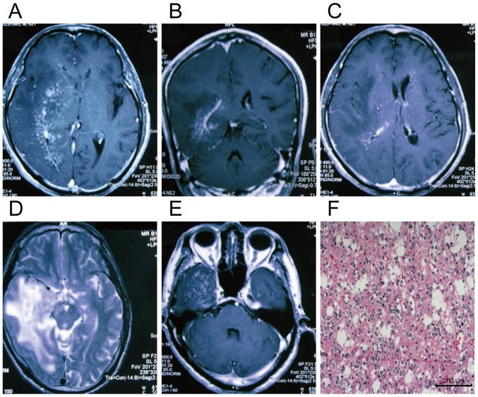
Figure 3.
MRI and pathological data of a glioma patient. Male patient aged 62 years. (A-E) The MRI scanning images in different levels, and (F) shows the postoperative pathological image of glioma in the same patient (magnification, ×200). (C) The image on T2WI sequence, and (A, B and D) T1WIs after enhancement. Large areas of irregular abnormal signals are visible in the right temporal, parietal and occipital lobes, basal ganglia and centrum semioval. There are mixed isointense and hypointense signals on the T1WI as well as mixed isointense and hyperintense signals on the T2WI, and the surrounding edemas are not enhanced. MRI, magnetic resonance imaging; T1WI, T1-weighted image.

