Fig. 1.

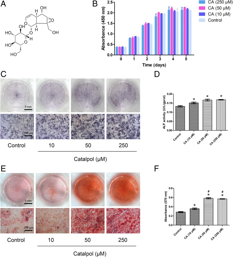
Catalpol promotes the osteogenic differentiation of BMSCs in vitro. a Chemical structure of catalpol. b Cell viability after catalpol treatment was determined using the CCK-8 assay. c–f Osteogenic differentiation was assessed by ALP staining (c), ALP activity assays (d) and Alizarin Red staining (e). Calcium deposition was determined by measuring optical density (f). The data were confirmed by three repeated tests. The data are presented as the means ± SD. CA, catalpol. *P < 0.05 compared with the control group, #P < 0.05 compared with the 10 μM catalpol treatment group
