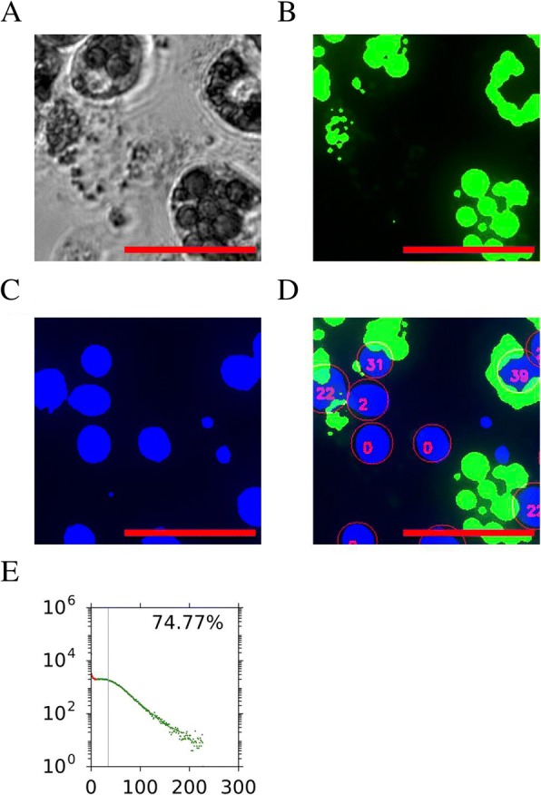
Fig. 2.

Representative analysis of 3T3-L1 cells after adipogenic stimulation. The representative phase-contrast image (a), binarised green lipid channel (b), and blue nuclei (c) channels are shown. The final calculated score for each of the detected nuclei is shown in d, where the number within each nucleus indicates the calculated lipid score for each of the nuclei. Note that blue-stained regions that were too small were excluded from the calculations. Finally, a histogram is created for all of the nuclei counted within the well, and the percentage of nuclei that have lipid scores above the threshold (in green dots) is indicated as adipogenic score in the upper right corner (e). For all the histograms, X-axis is the lipid score and Y-axis is the number of cells with the particular score. The vertical grey line indicates mean lipid fluorescence levels per nuclei for that cell population. Scale bars of images, 100 μm
