Fig. 5.
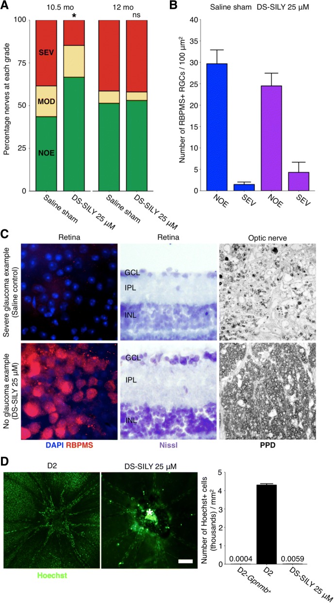
DS-SILY protects from D2 glaucoma at 10.5 mo of age. 25 μM DS-SILY was administered every 21 days via intravenous injection to the tail vein of D2 mice starting at 6–7 mo of age. Mice administered DS-SILY and saline sham controls were harvested at 10.5 and 12 mo and optic nerves and retinas were assessed for glaucomatous neurodegeneration. a DS-SILY administration significantly protected D2 eyes from glaucoma at 10.5 mo as assessed by optic nerve assessment (green = NOE; no detectable glaucoma but called no or early glaucoma as some eyes have early gene expression changes, yellow = MOD; moderate glaucoma, red = SEV; severe glaucoma). b Agreeing with this, RGC numbers were preserved in protected eyes with no detectable glaucoma (blue bars = saline sham, purple bars = DS-SILY). Eyes with severe glaucoma had also lost their RGCs indicating that DS-SILY treatment did not uncouple somal and axonal degeneration (SEV bars)). c Examples of no glaucoma and severe glaucoma retinas as assessed by RBPMS staining (an RGC marker; left; n = 5 for each condition), Nissl staining (center; n = 5 for each condition), and optic nerve cross sections assessed by PPD staining (right; n = 55 saline 10.5 mo; 64 saline 12 mo; 80 DS-SILY 10.5 mo; 66 DS-SILY 12 mo). Top row shows examples of severe retinas from representative eyes with severe optic nerve damage (SEV) as assessed by PPD staining of the optic nerve, bottom row shows examples of retinas with no neurodegeneration in the optic nerve (NOE graded nerves). d DS-SILY administration potently prevents vascular leakage in the retina at 9–9.5 mo (n = 18). Vascular leakage was assessed by an intravenous injection of the nuclear label Hoechst (green). Retinas were flat-mounted and Hoechst positive ganglion cell layer nuclei (excluding vascular endothelial cell nuclei) were counted across the retina from 8 representative regions in each eye. The bright region (*) over the optic nerve head in the right hand panel is due to labelling of cells in a residual tuft of hyaloid vasculature over the ONH (as often occurs in mice) and does not indicate increased leakage in the optic nerve. Scale bar = 100 μm. The numbers above D2-Gpnmb+ and DS-SILY 25 μM represent the data points

