Fig. 4.
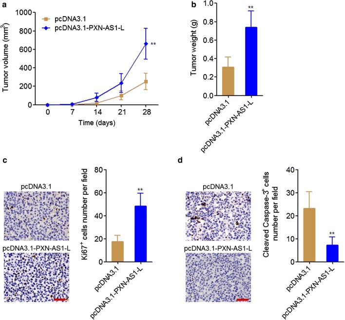
PXN-AS1-L promoted NSCLC xenograft growth in vivo. a Tumor volumes of subcutaneous xenografts derived from PXN-AS1-L stably overexpressed and control A549 cells were measured every 7 days. b Tumor weights of subcutaneous xenografts derived from PXN-AS1-L stably overexpressed and control A549 cells at the 28th day after injection. c In vivo cell proliferation of PXN-AS1-L stably overexpressed and control A549 cells was evaluated using Ki67 immunohistochemistry staining of subcutaneous xenografts. Scale bars = 50 µm. d In vivo cell apoptosis of PXN-AS1-L stably overexpressed and control A549 cells was evaluated using cleaved caspase-3 immunohistochemistry staining of subcutaneous xenografts. Scale bars = 50 µm. Results are shown as mean ± SD of six mice in each group. **P < 0.01 by Mann–Whitney test

