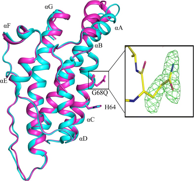
Fig. 5.

Ypd1-G68Q structure is similar to that of wild-type Ypd1. Overlay of wild-type Ypd1 (cyan) (PDB ID: 1QSP) and the Ypd1-G68Q mutant (magenta). The helices are numbered sequentially A to G from the N terminus to the C terminus, with the four-helix bundle core composed of helices B, C, D and G. The phosphorylatable histidine and H + 4 glycine or glutamine are shown in stick representation. Movement of the αA helix is observed with a RMSD of 1.7 Å for this region. Inset: Electron density for the substituted Q residue at position 68 in Ypd1 as shown by the Fo-Fc omit map (green mesh), contoured at 3.0 σ
