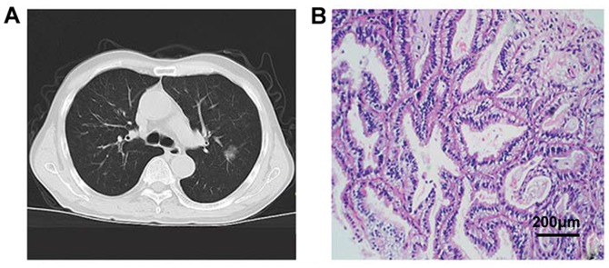
Figure 2.
CT signs and pathological data of GGN. (A) CT shows a GGN on the right upper lung with a diameter of ~8 mm and contains solid components. (B) H&E staining suggests epithelial adenocarcinoma, presence of alveolar structures, alveolar wall thickening, and cancer cells growing along the alveolar wall. GGN, ground-glass nodule.

