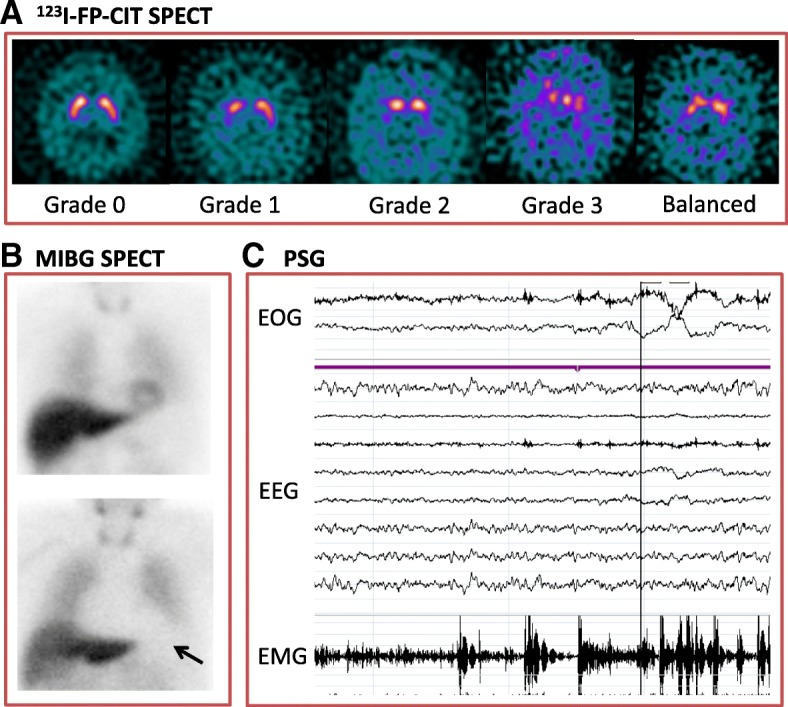
Fig. 1.

Indicative biomarkers for dementia with Lewy bodies. A. N-ωfluoropropyl-2β-carbomethoxy- 3β-(4-iodophenyl) nortropane (123I-FP-CIT SPECT) single photon emission tomography (SPECT). Axial images from FP-CIT SPECT at the level of the striatum. Grade 0 – normal uptake in left and right striatum. Grade 1 – unilateral decreased uptake in putamen [42]. Grade 2: bilateral uptake in putamen. Grade 3: virtually absent uptake bilaterally in the caudate and putamen. Balanced bilateral loss in the caudate and putamen is often seen in DLB, which does not fit easily into any Benamer scale category. B. Cardiac Meta-iodobenzylguanidine (MIBG SPECT) Imaging. The top image is normal, with a clear cardiac outline visible (arrow, HMR=3.14). The bottom image is abnormal with no visible cardiac outline (HMR=1.03). C. Polysomnography (PSG) recording demonstrating episodes of REM sleep without atonia on electro-oculogram (EOG) measuring eye movements, electroencephalogram (EEG) and electromyogram (EMG) measuring chin movement. With thanks to Dr Sean Colloby (a), Ms Gemma Roberts (b) and Dr Kirstie Anderson (c)
