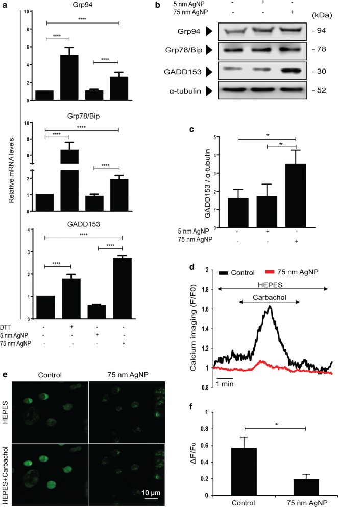
Fig. 5.
75 nm AgNP treatment depletes ER calcium stores and leads to ER stress. a Relative mRNA levels of ER stress markers in MCF-7/KCR cells treated with 75 nm AgNPs. b Protein levels of ER stress markers detected by immunoblot. c Densitometric quantitation of GADD153 protein levels. d Histogram of real-time calcium imaging from at least 5 ROIs (Region of interest) and e fluorescent calcium imaging of untreated and 75 nm AgNPs-treated MCF-7/KCR cells upon carbachol administration. Pictures were taken before and 1 min after carbachol exposure. f Representative bar graph of cytoplasmic calcium released on carbachol exposure. The values represent the mean ± standard deviation calculated from three independent experiments (*P < 0.03 ****P < 0.0001, Fisher’s LSD test)

