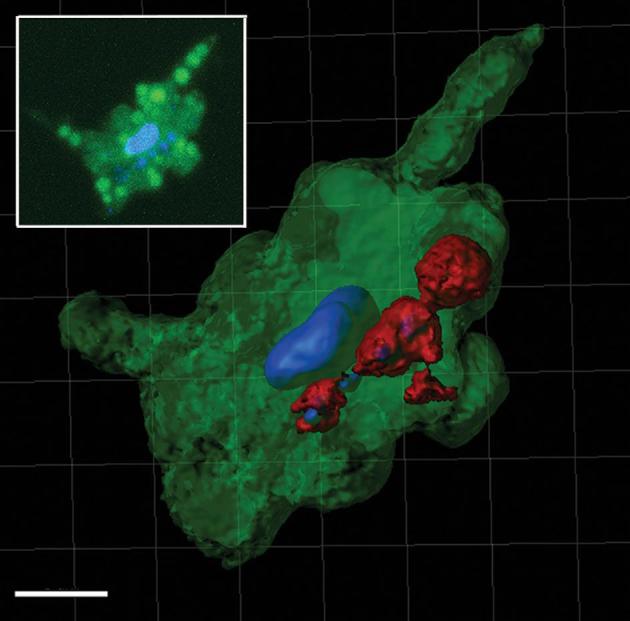
Figure 3.

Processed confocal z-stack image using the Contour Surface tool in IMARIS v.8.1 (Bitplane) of a DAPI stained H. hauckii-R. intracellularis symbiosis showing the proximity of the symbiotic trichome (red), stained nucleoids of symbiont (blue) and the diatom stained nucleus (blue). In green the diatom chloroplasts reconstruction and includes background. Scale bar 5 μm. Inset: parallel z-stack confocal image simultaneously excited by 405 and 488 laser lines highlighting the diatom nucleus and the cyanobacteria nucleoids (in blue), and chloroplasts (brighter green circles; note, a lower degree of averaging shows some background auto-fluorescence).
