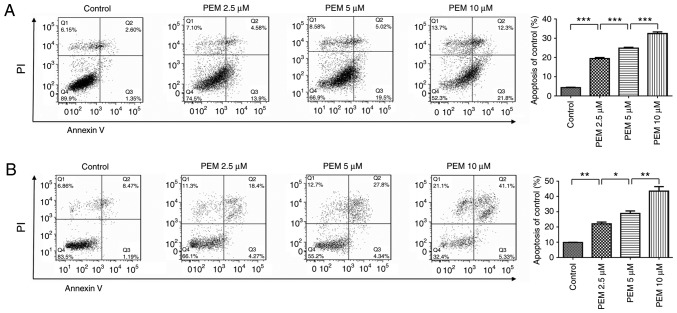
Figure 3.
Pemetrexed induces apoptosis in human esophageal squamous cell carcinoma cells. (A) Eca-109 and (B) EC9706 cells were treated with 0, 2.5, 5 or 10 µM pemetrexed and incubated for 36 h. Following treatment, the cells were harvested for apoptosis assays. The percentage of apoptotic cells was quantified using FlowJo software and analyzed using SPSS software. All data are presented as the mean ± standard deviation. *P<0.05, **P<0.01, ***P<0.001. Q1: (Annexin V− FITC)−/PI+, necrotic cells. Q2: (Annexin V−FITC)+/PI+, late apoptotic cells. Q3: (Annexin V− FITC)+/PI−, early apoptotic cells. Q4: (Annexin V− FITC)−/PI−, normal control cells. PI, propidium iodide; FITC, fluorescein isothiocyanate; PEM, pemetrexed.

