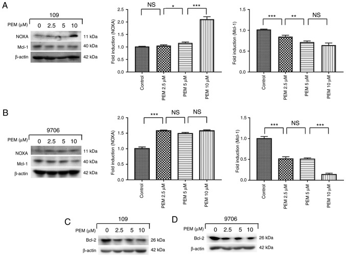
Figure 6.
Involvement of the NOXA/MCL-1 axis in pemetrexed-induced apoptosis. (A and C) Eca-109 and (B and D) EC9706 cells were treated with 0, 2.5, 5 or 10 µM pemetrexed and incubated for 36 h. Following treatment, NOXA and Mcl-1 expression was quantified via western blot analysis. All data are presented as the mean ± standard deviation. *P<0.05, **P<0.01, ***P<0.001. PEM, pemetrexed; NOXA, phorbol-12-myristate-13-acetate-induced protein 1; Mcl-1, induced myeloid leukemia cell differentiation protein Mcl-1; Bcl-2, apoptosis regulator Bcl-2; NS, not significant.

