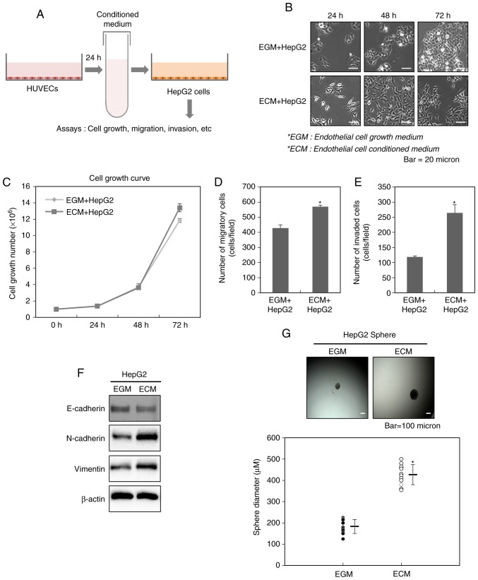
Figure 1.
Endothelial cells increase the malignant potential of liver cancer cells. (A) A schematic illustration showing the procedure for the acquisition of the conditioned medium obtained from HUVECs and the applications in this study. (B) Morphological changes of HepG2 cells after the treatment with ECM. (C) Effect of ECM on the growth of HepG2 cells. After HepG2 cells were treated with ECM, the cell counting was performed at 24, 48 and 72 h. Increased (D) migration and (E) invasion of HepG2 cells after the treatment with ECM. The migratory and invasive properties of the cells were measured using the Transwell chamber. (F) Western blot analysis for the expressions of N-cadherin and vimentin after the treatment with ECM. Experiments were performed in triplicate, and the data shown are representative of a typical experiment. (G) Quantification of sphere-forming abilities of HepG2 cells after the treatment with ECM. The cells were grown in DMEM/F12 supplemented with B27, N2, basic fibroblast- and epidermal growth factor onto 24-well ultra-low attachment plates at 300 cells per well for 7 days, and the size of spheres were determined. To measure the size of sphere, 12 spheres per group (n=12/group) were randomly selected. The average size of each sphere is quantified in the standard deviation and shown in the representative graph. Results from three independent experiments are expressed as mean ± 1 SEM (*P<0.05). HUVECs, human umbilical vein endothelial cells; ECM, endothelial cell culture medium.

