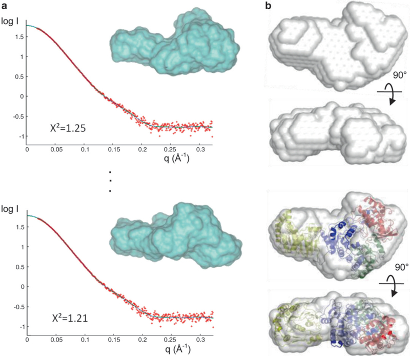
Fig. 3.

Applications of SAXS. (a) De novo shape determination using GASBOR. Ten individual runs generate ten different models (black lines) that fit the experimental scattering curve (red) well with X2 values between 1.2 and 1.4; two of the ten curves and de novo models (blue surface) are shown. (b) Two views of an averaged molecular envelope obtained from (a). In this example, the protein consists of two domains whose independent crystal structures are known. These structures were fitted into the SAXS envelope using SUPCOMB to generate a full-length model of the protein (bottom)
