Figure 4:
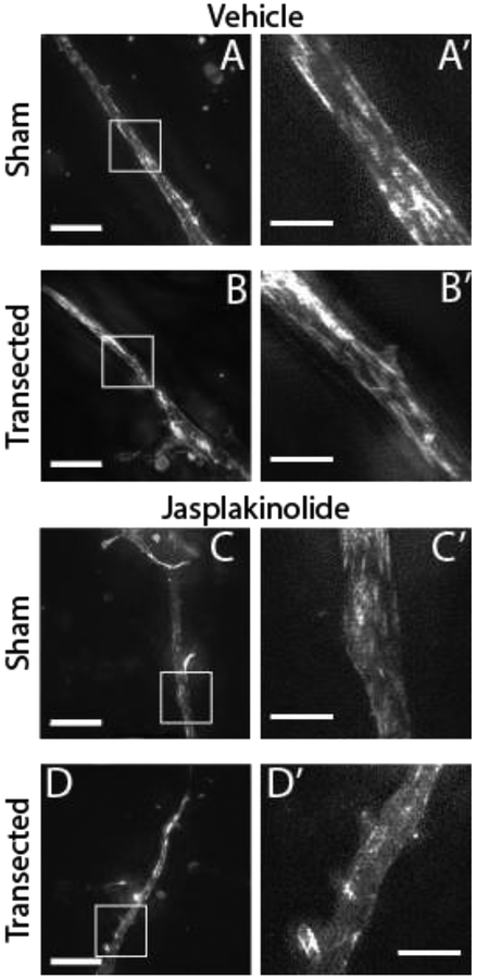
Neurites contain longitudinal bundles of F-actin that are diminished by jasplakinolide treatment but not by transection. (A-D) Maximum intensity projections of 3D-SIM images of neurites at the first time point (mean 2.5 min post transection, SD 1.5 min) stained with SiR-Actin, with and without transection and/or jasplakinolide treatment (scale bars: Left = 10 μm, Right = 3 μm). Note that the dynamic range of the image was optimized independently for each neurite.

