
Figure 3. Dopamine neuron subtype projections to the caudate putamen.
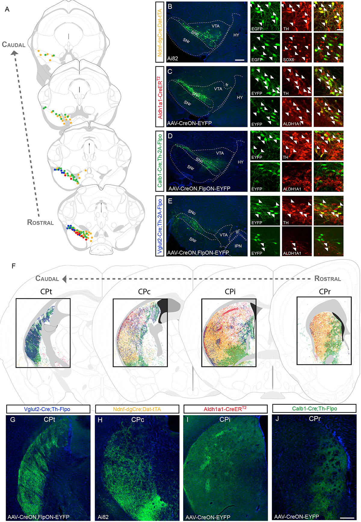
(A) Distribution of DA neuron subtypes used in the intersectional genetic experiments to trace projections in the caudate putamen (CP; each dot = 3 neurons; yellow = Ndnf experiment shown in B, red = Aldh1a1 experiment shown in C, green = Calb1 experiment shown in D, blue = Vglut2 experiment shown in E). (B) In Ndnf–dgCre;Dat–tTA;Ai82 mice, EGFP+ cell bodies are located throughout the SNc, the dorsolateral VTA and RR. The vast majority of these cells express TH and SOX6 (arrowheads). (C) Example of a Aldh1a1–CreERT2 brain injected in the SNc with AAV–CreON–EYFP virus. EYFP cells were positive for TH and ALDH1A1. (D) Injection of Calb1–Cre;Th–2A–Flpo mouse with a AAV–CreON,FlpON–EYFP virus in the SNc. Most EYFP+ cells did not express ALDH1A1. (E) Injection of a AAV–CreON,FlpON–EYFP virus in the SNc of Vglut2–Cre;Th–2A–Flpo mouse yields labeled neurons in the dorsolateral part of the SNc (corresponding in part to the pars lateralis). A small fraction of these neurons is ALDH1A1+ (arrowhead). (F) Distinctive areas of the dorsal striatum are innervated in the experiments described above, shown at different rostrocaudal levels (CPr = rostral, CPi = intermediate, CPc = caudal, CPt = tail). (G-J) Representative images from these experiments (See Table S1 for number of animal replicates for each experiment). Scale bars: B-E low mag. = 200 μm, high mag. = 40 μm; G-J =200 μm.