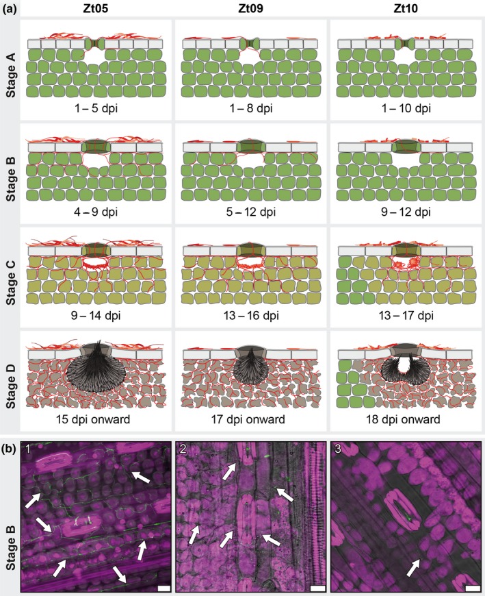
Figure 3.

Zymoseptoria tritici wheat infections are characterized by four distinct infection stages and isolate‐specific infection development. (a) Schematic drawings of the key features that characterize the four infection stages of Z. tritici and illustrate the infection phenotypes of isolates Zt05, Zt09, and Zt10 on the wheat cultivar Obelisk. (b) Micrographs showing Z. tritici hyphae (arrows) during biotrophic growth inside wheat leaves. Maximum projections of confocal image z‐stacks. Nuclei and wheat cells are displayed in purple and fungal hyphae or septa in green. The panel shows biotrophic colonization of (1) isolate Zt05 at 7 dpi, (2) Zt09 at 11 dpi, and (3) Zt10 at 9 dpi. Scale bars = 25 µm
