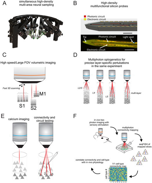
Figure 3: New methods for dissection cortical layer function.
A) Schematic of a mechanical setup for the simultaneous insertion of multiple ultra-high density multielectrode arrays (courtesy of New Scale Technologies). B) Schematic of a multi-layer silicon probe integrating electrodes and optical waveguides for recoding and optogenetics (courtesy of V. Lanzio and S. Cabrini). C) Schematic of a very large field of view high speed volumetric two photon microscope for densely imaging across multiple layers and multiple connected cortical areas simultaneously. D) Schematic of using a holographic multiphoton microscope to selectively perturb individual layers in the same animal on different trials. E) Schematic of a paradigm for mapping the physiological responses of L2/3 and L4 cortical neurons with calcium imaging, and then optogenetically stimulating a precise ensemble of presynaptic L4 neurons while recording from a L2/3 neuron in attempt to recapitulate the sensory response properties of the L2/3 neuron. F) Sequence of proposed experiments for correlating the sensory physiology, synaptic connectivity, and transcriptionally defined cell types of densely imaged cortical neurons across layers.

