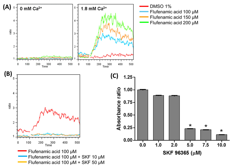
Figure 3. Induction of Ca2+ influx in MKs and effect of SKF96365 on MK cell proliferation.
(A) Ca2+ influx was determined by time-dependent flow cytometry using flufenamic acid (FFA) at different concentrations in day 12 MKs in Ca2+-free buffer (0 mM Ca2+), left, and in the presence of external Ca2+ (1.8 mM Ca2+), right. The change in fluorescence signals of the Ca2+-binding dyes, Fluo-4 and Fura Red was acquired in the FL-1 and FL-3 channels, respectively, and the ratio (FL-1/FL-3) is presented as a function of time. The curves are representative of three independent experiments. (B) MK cells at day 12 were pre-incubated with SKF96365 at 10 and 50 μM for 3 min and then activated with 100 μM FFA in the presence of external Ca2+. (C) MK cells on day 10 were cultured for 48 h in the presence of 50 ng/mL TPO with increasing concentrations of SKF96365. Cell proliferation was determined using the MTT assay. Data shown are the mean ± SD of triplicate measurements compared to the control, 0 μM SKF96365. * P-value < 0.05.

