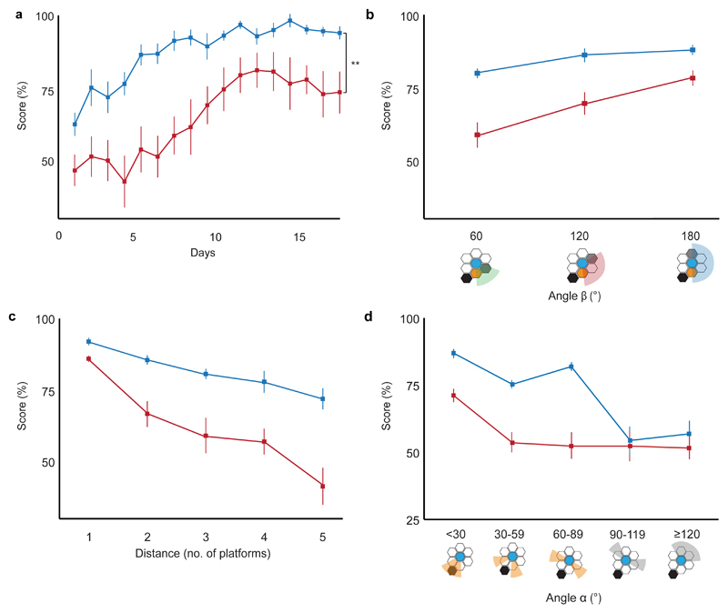
Figure 4. Hippocampal-lesioned animals on the Honeycomb Maze.
(a) Animals with hippocampal damage (red, n = 8) are significantly impaired in learning the task compared to operated controls with sham hippocampal lesions (blue, n = 8) (F1,14 = 10.240, p = 0.006, two-way Mixed ANOVA), 4 trials per day. (b-d) Animals with hippocampal damage (red, n = 8) are significantly more influenced than controls (blue, n = 8) by (b) separation between choice platforms (angle β; F2,28 = 40.024, p < 0.001, two-way Mixed ANOVA), (c) distance from the goal (F4,56 = 34.740, p < 0.001, two-way Mixed ANOVA), and (d) angle to goal (angle α; F2.3, 32.8 = 28.812, p < 0.001, two-way Mixed ANOVA). For (a-d) error bars indicate s.e.m., ** p < 0.01.

