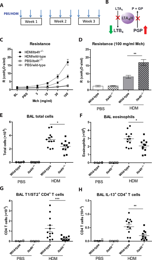
Fig. 1. Augmented airway resistance in HDM-treated lta4h−/− mice despite reduced airway inflammation.
(A) Wild-type and lta4h−/− mice were administered HDM or phosphate-buffered saline (PBS) intranasally three times per week for 3 weeks. (B) Schematic depicting the opposing proinflammatory (LTB4 generation) and anti-inflammatory (PGP degradation) activities of LTA4H. Abrogation of LTA4H activity (red crosses) will result in reduced LTB4 but increased PGP. (C) Airway resistance (R) to increasing doses of methacholine (Mch) was measured 24 hours after the final HDM/PBS exposure. (D) Airway resistance at 100 mg/ml methacholine. At 24 hours after final HDM/PBS exposure, total cell numbers in the BAL were assessed by trypan blue exclusion (E), and airway eosinophils (F), CD4+ T cells expressing T1/ST2 (G), and CD4+ T cells expressing IL-13 (H) were assessed by flow cytometry. Figures present combined data from two independent experiments with four to six mice per group in each experiment. Results depicted as means ± SEM. *P < 0.05, **P < 0.01, ***P < 0.001, using Mann-Whitney statistical test.

