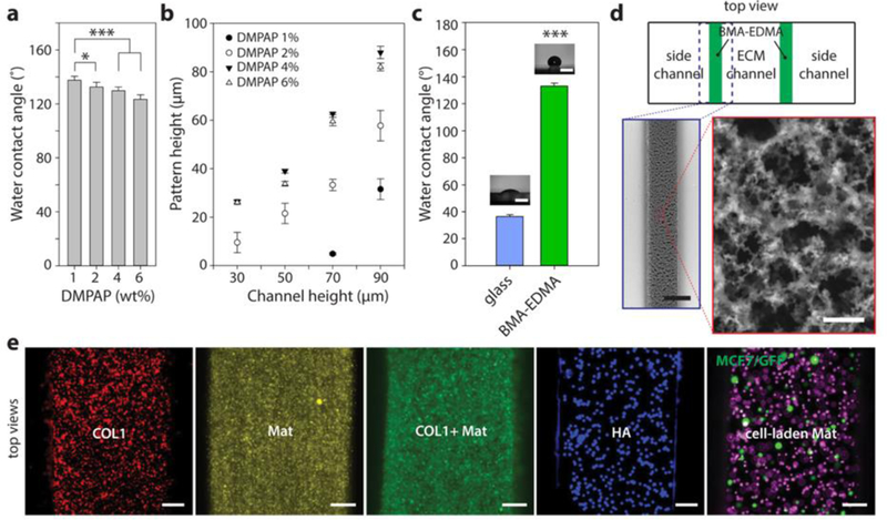
Figure 2. Characterization of BMA-EDMA and hydrogel patterning within the microfluidic device.

a. Water contact angle (WCA) of hydrophobic pattern as a function of the concentration of photoinitiator, 2,2-dimethoxy-2-phenylacetophenone (DMPAP), in the BMA-EDMA solution. b. Resulting height of polymerized BMA-EDMA patterned using primary PDMS slabs with four different channel heights at four different DMPAP concentrations. n=3. c. Average WCAs of a bare glass coverslip and BMA-EDMA pattern (1% DMPAP, 70 µm channel height for pattern). Images show representative WCA measurements for each condition. Scale bars: 200 µm. ***p<0.001. n=3. d. (counterclockwise from top) Schematic top view of the BMA-EDMA patterned device (not to scale). Scanning electron microscope (SEM) images of the BMA-EDMA pattern show the microporous structure of the pattern which contributes to the hydrophobic property of the pattern. Scale bars: 250 µm (bottom left) and 10 µm (bottom right). All error bars: one standard deviation. e. A variety of hydrogel types, including COL1, MAT, COL1/MAT (1:1 volume ratio), HA, and MCF-7/GFP breast cancer cell-laden MAT were injected into the hydrogel channel of the BMA-EDMA device to demonstrate hydrogel filling. To visualize hydrogels in the device, fluorescent microspheres (5–14 µm; Spherotech, USA) were mixed in each gel solution at a volume ratio of 10 : 1 (hydrogel : DPBS containing fluorescent microsphere (1.0% w/v)). All hydrogels, from the less viscous COL1 (2 mg/mL) to the highly viscous HA (4%), were successfully filled and polymerized without overflowing over the hydrophobic pattern. Scale bars: 100 µm.
