
Figure 3. 3D breast epithelial cancer cell migration through a fibrous structural extracellular matrix (ECM) and a mixture of fibrous structural ECM and adhesive proteins within the BMA-EDMA device.
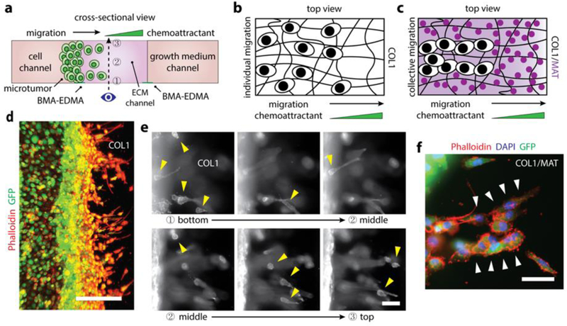
a. Schematic cross-sectional view of the 3D breast epithelial cancer cell migration assay. Two ECM structures, COL1 and COL1/MAT, were formed in the ECM channel (center) created by the BMA-EDMA pattern. MCF-7/GFP cells were seeded in the cell channel (left) to form a 3D microtumor tissue. A complete growth medium containing 5% fetal bovine serum (FBS) was filled into the cell channel after cell attachment, and a complete growth medium containing 10% FBS was filled into the growth medium channel (right). Dotted arrow indicates the imaging axis for the assay. Numbers indicate focal planes corresponding to the images shown in (e), from bottom (1) to top (3). b. An illustration (top view) for the migration behavior of MCF-7/GFP cells within the COL1 fibrous structural ECM. c. Illustrative top view of MCF-7/GFP cells migrating through the COL1/MAT mixture of fibrous structural ECM and adhesive proteins. d. Fluorescent image of the MCF-7/GFP cells (on day 4) migrating through COL1. Actin filaments of the MCF7/GFP cells were immunostained with rhodamine Phalloidin (red) and the cells were stably transfected to express GFP. Scale bar: 300 µm. e. Fluorescent images corresponding to the focal planes in (a), from bottom (1) to top (3). Yellow arrow heads indicate the migrating cells in each focal plane, indicating that the ECM environment created by the BMA-EDMA hydrophobic pattern is truly three-dimensional. f. A fluorescent image shows collectively migrating MCF-7/GFP cells (on day 4) through COL1/MAT as shown in (c), in contrast with COL1 alone, in which cells migrated individually as shown in (b). White arrowheads indicate collectively migrating MCF7/GFP cells. Actin filaments (red) and nuclei (blue) were immunostained with rhodamine Phalloidin and 4’, 6-diamidino-2-phenylindole (DAPI), respectively. Scale bar: 100 µm.