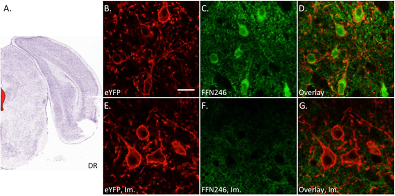
Figure 5.
FFN246 labels 5-HT neurons in the mouse dorsal raphe in an mSERT-dependent manner. A) Mouse brain atlas image highlighting the dorsal raphe (DR) region containing the 5-HT neurons (Bregma: −4.5 mm, Allen Brain Institute).51 Acute brain slices were collected from Pet1-cre/flox-ChR2-eYFP mice and incubated with FFN246 (20 μM) for 30 min. Uptake of FFN246 (green) in eYFP-positive neurons (red) was counted where FFN signal was greater than two standard deviations above background. 75% of eYFP neurons loaded FFN246 (81/108 neurons from 4 animals). Representative images: eYFP (B), FFN246 (C), and overlay (D). Preincubation with imipramine (Im., 2 μM) completely eliminated FFN246 uptake in 5-HT neurons (0/49 neurons from 3 animals). Representative images of SERT inhibited conditions: eYFP (E), FFN246 (F), and overlay (G). Scale bar, 20 μm.

