Figure 6.
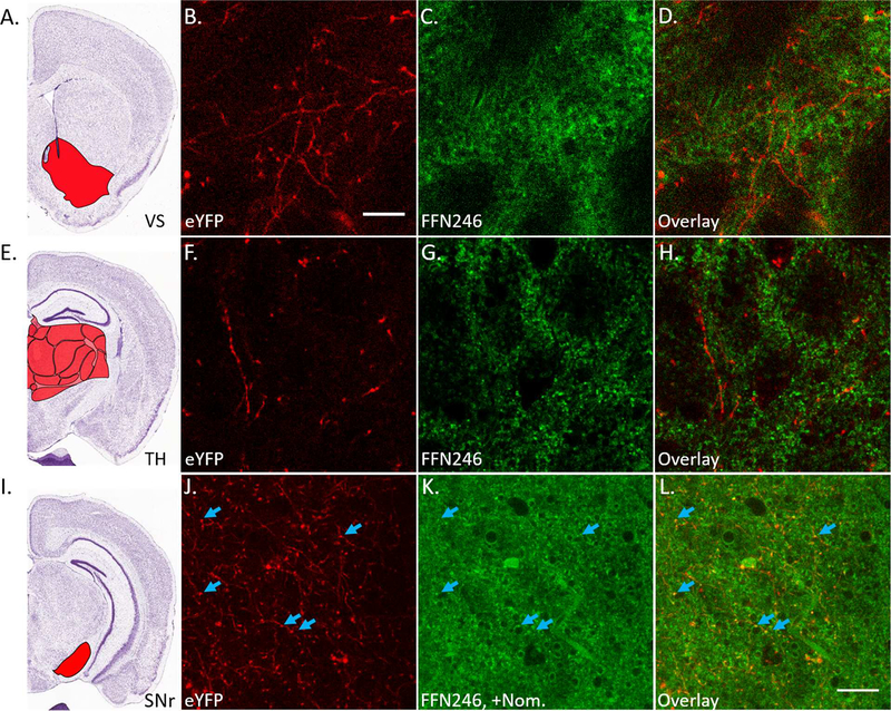
FFN246 does not significantly label 5-HT axonal projections. A) Mouse brain atlas image highlighting the ventral striatum (VS, Bregma: +0.5 mm, Allen Brain Institute).51 Brain slices were collected from Pet1-cre/flox-ChR2-eYFP mice and incubated with FFN246 (20 μM) for 30 min. There was no observable colocalization between eYFP (red) and FFN246 (green) in 5-HT neuronal projections (i.e. no FFN signal greater than 2SDs above background in eYFP-positive strands from 3 animals). Representative images: eYFP (B), FFN246 (C), and overlay (D). E) Mouse brain atlas image highlighting the thalamus (TH, Bregma: –1.5 mm).51 Again, no colocalization between eYFP and FFN246 was observed (3 animals). Representative images: eYFP (F), FFN246 (G), and overlay (H). I) Mouse brain atlas image highlighting the substantia nigra reticulata (SNr, Bregma: −2.5 mm).51 In this brain region, high FFN246 background staining was also observed even with preincubation of nomifensine (2 μM). However, some puncta colocalized with eYFP axonal signal (blue arrows). Scale bar, 20 μm.

