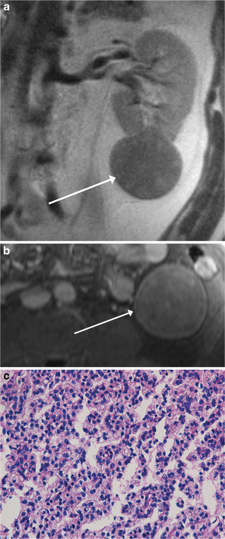
Fig. 1.
MR images of a 51-year-old man with a renal mass. Coronal single-shot T2-weighted image (a) and axial post-contrast fat-suppressed T1-weighted image (b) show a well-encapsulated T2-hypointense solid mass extending exophytically from the left lower pole (arrow). The mass showed diffuse low-level internal enhancement on subtracted post-contrast images (not shown). The findings were consistent with MRI appearance I. Mass was diagnosed as type 1 papillary renal cell carcinoma (pRCC) at partial nephrectomy and was therefore classified as focal type 1 pRCC in our study. Section of tumour (c) shows a relatively solid area of tumour with small neoplastic nuclei without prominent nucleoli, indicative of low nuclear grade (H&E stain, ×400); overall, the lesion was classified as being of predominantly low nuclear grade, with high nuclear grade present (not shown). On follow-up cross-sectional imaging 7.0 years later, there was no evidence of metastatic disease

