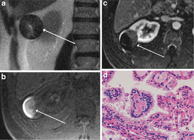
Fig. 2.
MR images of a 55-year-old man with a renal mass. Axial single-shot T2-weighted image (a), axial pre-contrast fat-suppressed T1-weighted image (b) and subtracted axial post-contrast fat-suppressed T1-weighted image (c) show a heterogeneous, predominantly T2 hypointense mass, extending exophytically from the lateral right kidney (arrow, a). There is T1 hyperintensity within the lateral aspect of the mass (arrow, b), indicative of haemorrhagic content, as well as low-level enhancement within the medial non-haemorrhagic portion of the mass (arrow, c), findings consistent with MRI appearance II. Mass was diagnosed as type 2 papillary renal cell carcinoma (pRCC) at partial nephrectomy and was therefore classified as focal type 2 pRCC in our study. Section of the tumour (d) shows papillary fronds lined by neoplastic cells with small nuclei (centre of the field), indicative of low nuclear grade, compared with a minor component on the right-hand side of the image with larger nuclei and conspicuous nucleoli, indicative of focal high nuclear grade (H&E stain, x400); overall, the lesion was classified as predominantly low nuclear grade, with high nucleargrade present. On follow-up cross-sectional imaging 4.9 years later, there was no evidence of metastatic disease

