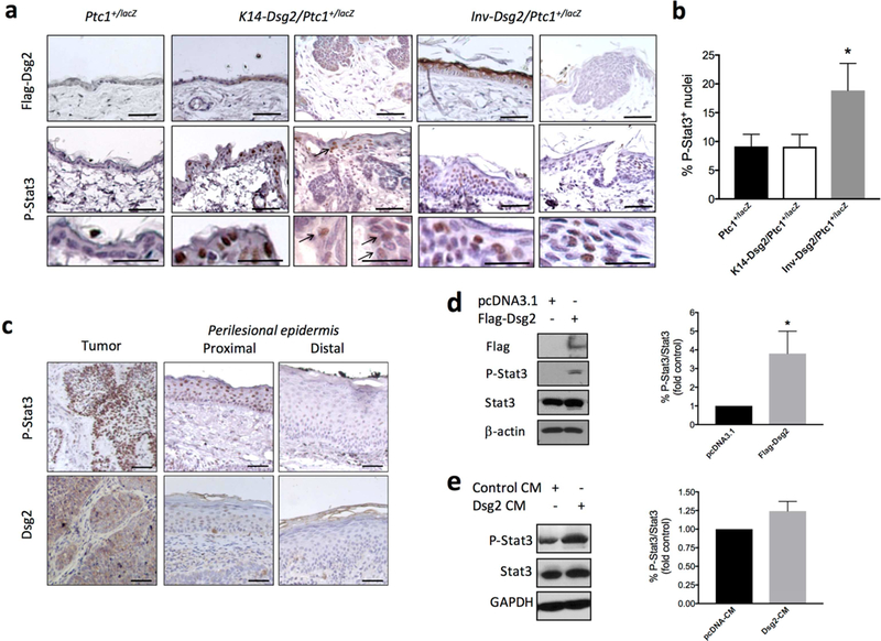
Figure 2. Dsg2 increased Stat3 phosphorylation in murine and in human sporadic BCCs.

(a) Formalin-fixed, paraffin-embedded skin sections from 6-month old Ptc1+/lacZ and Inv-Dsg2; Ptc1+/lacZ and 3-month old K14-Dsg2/Ptc1+/lacZ mice were immunostained for Flag (top panels), and P-Stat3 (bottom panels). Scale bars = 50 microns, inset scale bar = 100 microns. Representative images from n=3–5 animals. (b) Quantification of positive and highly positive P-Stat3+ nuclei in the interfollicular epidermis (not tumor areas) of the indicated mouse genotypes. Graph represents mean ± SEM (n=3–5; *P<0.05) (c) Representative example of human BCC immunostained for Dsg2 and P-Stat3. IHC staining of tumor, and both proximal and distal perilesional skin are shown. (d) Representative Western blot analysis of Dsg2.Flag, P-Stat3 (Tyr705), Stat3, and β-actin in ASZ001 transfected with empty plasmid (pcDNA3) or with pcDNA3 encoding Flag-tagged Dsg2 (mDsg2) (n=3; *P<0.05) (e) Naïve ASZ001 were stimulated for 30 min with conditioned medium from cells transfected as in (d), followed by lysis and western blot analysis of P-Stat3(Tyr705) and total Stat3 levels (n=3; P=0.068).
