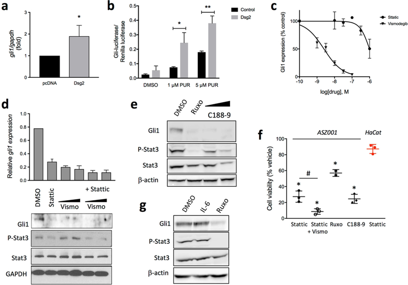
Figure 3. Stat3 mediates Dsg2-induced Gli1 upregulation and is essential for BCC cells viability.

(a) gli1 mRNA expression by qPCR of ASZ001 cells transiently transfected with empty plasmid (pcDNA3) or with pcDNA3 encoding Flag-tagged Dsg2 (mDsg2) (n=3) *p<0.05; Student’s t-test. (b) NIH3T3-LIGHT2 cells stably transfected with empty vector (black) of a pcDNA-Dsg2 (grey) and serum starved for 24 h in the presence of the Smo agonist purmorphamine (PUR). Representative results of Gli-luciferase activity (normalized to Renilla luciferase) from an experiment performed in triplicate (n=3). * P<0.05 **P<0.01, one-tailed Student’s t-test. (c) Inhibition of gli1 expression, by qPCR, in ASZ001 cells treated with increasing concentrations of Stattic or vismodegib for 24 h (n=3). (d) qPCR and Western blot analysis of Gli1 expression in cells treated with 1 μM Stattic, 50–100 nM vismodegib, or a combination of both for 48 h. All treatments are significantly reduced compared to control (p<0.001). n=3, *p<0.05, Student’s t-test. Efficacy of Stattic is shown below by changes in P-Stat3 (Tyr705). (e) Western blot analysis of Gli1 expression and Stat3 phosphorylation in ASZ001 cells treated with 20 μM ruxolitinib (Ruxo) or 1–3 μM C188–9 for 48 h. (f) Gli1 expression and Stat3 phosphorylation in ASZ001 cells stimulated with 10 ng/ml IL-6 or IL-6 plus 20 μM ruxolitinib (Ruxo) (n=3). (g) ASZ001 cells were incubated for 48 h with 1 µM Stattic, 1 µM Stattic plus 100 nM vismodegib, 20 μM ruxolitinib, or 3 μM C188–9. Cell viability was determined with the WST-1 assay and expressed as % of viability of cells treated with DMSO (vehicle). Student’s t-test, #p<0.05; *p<0.0001. In red, viability of HaCaT cells incubated with 1 µM Stattic for 48 h (n=3–4).
