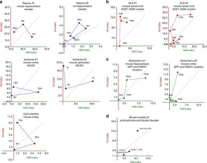
Fig. 4.
Time-dependent changes in the hiM- and hiI-indexes in animals subjected to various putative risk events for neuropsychiatric disorders and in genetic mouse models of schizophrenia, bipolar disorder, ALS, and Alzheimer’s disease. a Pattern of changes in the hiM- and hiI-indexes in the mouse and rat hippocampus after treatment with kainite (seizure #1 (GSE1831) and seizure #2 (GSE4236)), in mouse cortex and astrocytes after middle cerebral artery occlusion (MCAO; ischemia #1 (GSE32529), ischemia #2 (GSE35338)), and in mouse cortex after CpG infection (GSE32529). b Pattern of changes in the hiM- and hiI-indexes of the spinal cord of an ALS mouse model with the SOD1(G93A) mutation. c Pattern of changes in the hiM- and hiI-indexes of the hippocampus and cortex of an Alzheimer’s disease mouse model with mutations in APP and PSEN1. d hiM- and hiI-indexes in mouse models of schizophrenia and bipolar disorder

