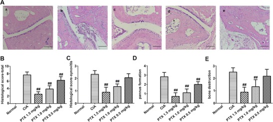
Fig. 2.

Histological analysis of inflamed joints in CIA mice. a The histological analysis was performed using mouse hind paw sections stained with HE (scale bars 100 μm): a normal group, b CIA group, c PTX 1.5 mg/kg group, d PTX 1.0 mg/kg group, and e PTX 0.5 mg/kg group. b–e Mean ± SEM histological arthritis scores (synovitis, pannus formation, bone destruction, and total) in the normal control group, CIA group, and three PTX treatment groups. #p < 0.05, ##p < 0.01 compared with the CIA group
