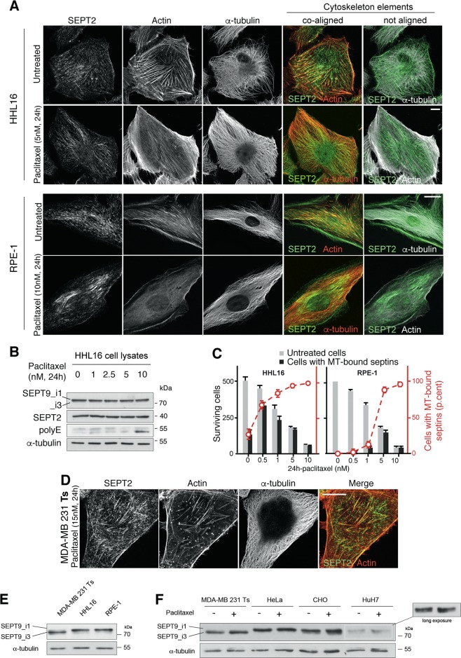
Fig. 7. Cells that survived acute paclitaxel treatment exhibit subcellular relocalization of endogenous septin filaments to microtubules.
a Immunofluorescent images of endogenous SEPT2 (green), actin (phalloidin; red or white) and α-tubulin in HHL16 and RPE-1 cells following 24 h paclitaxel treatment. The overlay images show that septin coalign with actin fibers in untreated cells, and with tubulin in paclitaxel treated cells. The paclitaxel concentration used corresponds to three times the IC50 for the drug. b Septin cellular amount does not change upon treatment with increasing concentrations of paclitaxel. Note that tubulin polyglutamylation starts increasing only after 24 h of 10 nM paclitaxel treatment. c Quantification of cells with MT-bound septins among cells that have survived 24 h-paclitaxel treatment. d Immunofluorescent images of breast cancer MDA-MB 231 cells after 15 nM paclitaxel treatment for 24 h. Note that septin filaments are still mainly associated with actin fibers in these cells. e Immunoblot showing the basal levels of SEPT9 long isoforms (SEPT9_i1 and _i3) in total cell extracts of untreated sensitive MDA-MB 231 Ts, HHL16, and RPE-1 cells. f Immunoblot showing the basal levels of SEPT9_i1 and _i3 in total cell lysates of untreated or paclitaxel-treated MDA-MB 231 Ts, HeLa, CHO, and HuH7 cells. The images, immunoblots shown and cell quantifications are representative of at least three independent experiments. Scale bars = 10 µm

