Fig. 3.
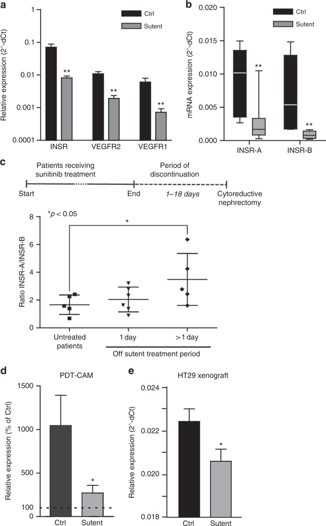
Altered INSR expression after angiostatic treatment with sunitinib. a, b INSR, VEGFR1 and VEGFR2 expression analysis in human RCC after treatment with sunitinib indicates a reduction expression of these growth factor receptors (A). Both INSR-A and INSR-B show reduced expression (b) **P < 0.01 by t test, N = 8–12, Ctrl vs. sunitinib. (c) Ratios of INSR-A vs. INSR-B expression in RCC. Surgery was performed in naive (untreated) patients, or after 1 or more (6–18) days of recovery from 2 cycles of sunitinib treatment prior to surgery. Prolonged discontinuation of sunitinib therapy results in induction of relative INSR-A levels, suggestive of rebound angiogenesis. *P < 0.05 by Mann–Whitney, N = 5. d qPCR profiling of the CAMs treated with PDT. Results are expressed as % of expression in untreated CAMs. The control bar (Ctrl) represents the increase in INSR expression by the PDT treatment, while the bar labelled sutent represents the normalisation of expression by sutent (20 μg/kg). Both bars present values of the situation 48 h post-PDT. *P < 0.05 (by t test). e qPCR analysis of HT29 CRC xenografts in mice treated with sunitinib,34 indicate that angiostatic treatment reduces vascular INSR expression. Special specific mouse primers identified INSR in mouse stromal tissue exclusively

