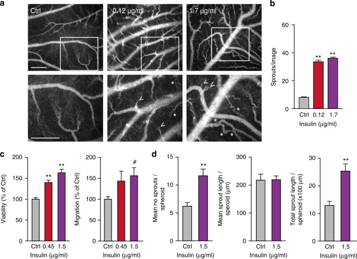
Fig. 4.
Insulin activates in vivo angiogenesis and in vitro endothelial cell functions. a Activation of angiogenesis in vivo in the chorioallantoic membrane (CAM) model. Representative fluorescence angiographies of the CAMs treated topically with various doses of insulin. CAMs were treated topically on development day 7 and 8 (20 μl in 0.9% NaCl) and visualised after i.v. injection of FITC-dextran (10 μl, 20 kDa) on day 9. Scale bar represents 100 μm. Arrow heads show new sprouting from pre-existing vessels, as quantified in (b). **P < 0.01 by ANOVA with Bonferroni’s Multiple Comparison test, N = 4. Asterisks indicate intussusceptive angiogenesis (not quantified). c Viability (left panel) and migration (right panel) of HMEC are stimulated by incubation with insulin for 72 h in medium containing reduced serum (0.5%). **P < 0.01 by ANOVA with Bonferroni’s Multiple Comparison test, N = 11; #P = 0.0657 by ANOVA with Bonferroni’s Multiple Comparison test, N = 5. d Effect of insulin on endothelial sprout formation by HUVEC in vitro. After embedding of spheroids in a 3D collagen gel, the number of sprouts per spheroid (left panel) and total sprout length (right panel) were significantly increased by treatment with insulin. However, mean sprout length (middle panel) did not change. **P < 0.001 by ttest, N = 6

