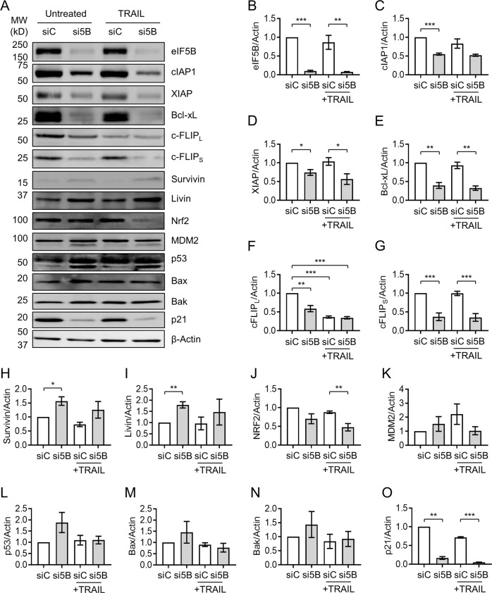
Fig. 5. Depletion of eIF5B leads to decreased levels of antiapoptotic proteins, p21, and Nrf2.
Control or eIF5B-depleted cells were treated, and protein harvested for immunoblotting, as in Fig. 4b (i.e., 100 ng/mL TRAIL for 4 h). a Representative images of immunoblots probing for eIF5B, cIAP1, XIAP, Bcl-xL, c-FLIPL, c-FLIPS, Survivin, Livin, Nrf2, MDM2, p53, Bax, Bak, p21, or β-actin (internal control). b–o Quantitation of eIF5B b, cIAP1 c, XIAP d, Bcl-xL e, c-FLIPL f, c-FLIPS g, Survivin h, Livin i, Nrf2 j, MDM2 k, p53 l, Bax m, Bak n, and p21 o, all normalized to β-actin. For p53 l, the top band (53 kDa) was quantified. Data are expressed as mean ± SEM for three independent biological replicates, except for Nrf2 (four biological replicates). *, p < 0.05; **, p < 0.01; ***, p < 0.001

