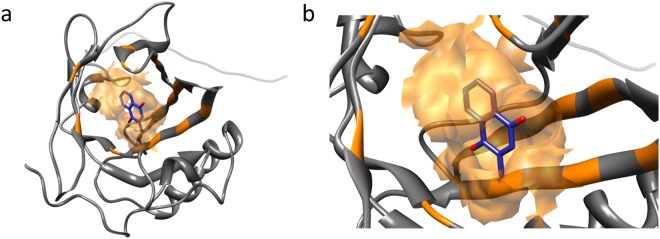
Figure 1.
Three-dimensional structure of murine MUP20 with menadione inside the binding cavity. (a) Inside the binding pocket, the ligand structure is depicted in blue with oxygen in red; the binding cavity surface and the interacting residues are highlighted in orange. The ligand is buried within the binding cavity and appears partially covered by the binding surface. (b) A closer view of the binding pocket reveals that the aromatic ring and the methyl group of menadione are inside the cavity. Docking was performed with Autodock/Vina42,43, starting from the first conformer of the NMR structure of MUP20 (2L9C.pdb).

