Figure 3.
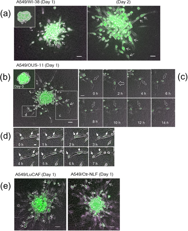
Collagen gel invasion of A549 cells (green) from spheroids with different types of fibroblasts. (a) A549/WI-38 spheroid incubated for 22 h (day 1) (left panel) and 44 h (day 2) (right panel). Inset, the original spheroid at day 0. (b) A549/OUS-11 spheroid incubated for 22 h (day 1). Inset, day 0. (c) Time-lapse images of the box “c” in (b) Arrows trace one cancer cell migrating on four fibroblasts in sequence. See Video 4. (d) Time-lapse images of the box “d” in (b) after 25-h incubation. Arrows trace two cancer cells showing reciprocating movement on a string of fibroblast. (e) A549/LuCAF (left panel) and A549/Ctr-NLF (right panel) spheroids after 22-h incubation (day 1). Note that both LuCAF and Ctr-NLF migrate very actively in the collagen matrix compared with WI-38 (a) and OUS-11 (b) at Day 1. Scale bars, 50 μm, except for 20 μm in (d) In all images, small arrows point cancer cells binding to fibroblasts.

